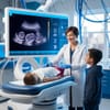
Researchers and clinicians are increasingly turning to artificial intelligence to improve how pediatric ultrasound images are captured, interpreted, and used for diagnosis. AI systems have shown promise in reducing user variability and increasing the accuracy of scans, particularly in settings where operators may not have deep expertise. For example, AI tools can assist novice users in acquiring high-quality lung ultrasound views, helping detect conditions like pneumonia with strong accuracy and efficiency.
In neonatal care, advanced AI models are being trained to interpret lung ultrasounds and predict critical respiratory outcomes. One study developed a neural network that analyzes early neonatal lung scans to estimate the need for respiratory support, such as CPAP, or treatments like surfactant — and the model achieved high predictive performance in clinical testing.
AI is also making strides in cardiac imaging for the very young. In pediatric and fetal echocardiography, machine learning and deep learning systems are helping automate the segmentation of heart structures, detect congenital heart disease, and perform quantitative measurements. These capabilities can lead to faster and more reliable diagnostics, especially in regions lacking highly specialized sonographers.
Beyond improving diagnostics, AI-driven “radiomics” is being used to extract rich, high-dimensional features from pediatric ultrasound images. By analyzing subtle textural and structural patterns beyond what the human eye can see, these radiomic models can help detect early, subclinical diseases — potentially transforming how pediatric pathologies are identified and treated.


